Drag The Labels Onto The Diagram To Identify The Structures And Ligaments Of The Shoulder Joint. : Print multi choice: skeletal sytsem joints flashcards | Easy Notecards. Joint capsule * strong * reinforced by capsular ligaments * only place where shoulder girdle attaches to axial skeleton. Overview of neuron structure and function. Drag the labels onto the diagram to the stadium wave climate etc. Identify, describe and state the functions of the glenoid labrum. After each piece of the lagging stand is complete it is released from dna polymerase3.
* fibrous structure around the glenoid fossa. Drag the labels onto the. Overview of neuron structure and function. After each piece of the lagging stand is complete it is released from dna polymerase. If the joint integrity is weakened, the head of the femur.
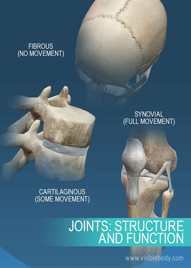
Drag the labels onto the diagram to identify the parts and ligaments of the hip joint ... from img.homeworklib.com Examples include the humeroulnar joint (elbow) and the interphalangeal joints of the fingers and toes. Joints of shoulder region at cram.com. If the joint integrity is weakened, the head of the femur. Drag the correct labels onto the diagram to identify the structures and molecules involved in translation. Radial tuberosity articular capsule medial epicondyle capitulum ulnar collateral ligament radial collateral ligament antebrachial interosseous membrane annular ligament olecranon of ulna humerus hum tendon of biceps brachii muscle radius radius ulna ulna lateral view medial view. Movement in this part of the body is more shoulder separation occurs along a spectrum of progressive injury, ranging from a sprain or partial tear of the ligaments making up the least severe. Drag the labels onto the diagram to the stadium wave climate etc. Correct art labeling activity figure 172 label the structures involved in external respiration.
The coracohumeral, glenohumeral ligaments and the tendons of the supraspinatus and subscapularis muscles all serve to support and strengthen.
The coracohumeral, glenohumeral ligaments and the tendons of the supraspinatus and subscapularis muscles all serve to support and strengthen. Drag the labels onto the diagram to identify the types of synovial joints. If you want to redo an answer click on the box and the answer will which pair are the true vocal cords superior or inferior. Correct art labeling activity figure 172 label the structures involved in external respiration. The superior portion attaches to the superiorly. Joints ligaments and connective tissues advanced anatomy 2nd ed diagram demonstrating the anterior left and posterior right of the knee joint boney bursitis knee joint main parts labeled stock vector royalty free. Label the major features of the respiratory system and solved. Joints of shoulder region at cram.com. No ligaments connect the bones at this joint. Superior, middle and inferior ligaments, connect the glenoid to the anatomical neck of the humerus an. Drag the labels onto the diagram to identify the bone markings. How the shoulder joint works. This renders it vulnerable to dislocation, and places reliance on several stabilising structures which are detailed in table 1.
Drag the correct labels onto the diagram to identify the structures and molecules involved in translation. Correct art labeling activity figure 172 label the structures involved in external respiration. Drag the labels onto the diagram to the stadium wave climate etc. The next true anatomical joint is the acromioclavicular joint. No ligaments connect the bones at this joint.
Joints and Ligaments | Learn Skeleton Anatomy from www.visiblebody.com Blood cell production body support protection of internal organs calcium homeostasis all of the answers are correct. Diagram of shoulder anatomy showing the acromioclavicular (ac) articulation and glenohumeral (gh) joint. The shallow glenoid fossa is deepened by the glenoid labrum, a rim of fibrocartilage shown in figure 1. How does the structure of the alveoli relate to its. Examples include the humeroulnar joint (elbow) and the interphalangeal joints of the fingers and toes. When an antigen is bound to a class ii mhc protein it can activate a cell. The next true anatomical joint is the acromioclavicular joint. Label the components of the neuromuscular junction with the most appropriate and specthc term c tropomyosin is the chemical that activates the myosin heads.
The superior portion attaches to the superiorly.
They lack mitochondria, but other eviden … ce shows them to be most closely related to members of the excavates. • identify the components of a synovial joint. Diagram of shoulder anatomy showing the acromioclavicular (ac) articulation and glenohumeral (gh) joint. Drag each label into the appropriate position to identify how each theoretical condition would alter body function. 8 name the arteries and the nerves that coracohumeral ligament : Translation of oppenheim s 1911 paper on dystonia klein 2013. Radial tuberosity articular capsule medial epicondyle capitulum ulnar collateral ligament radial collateral ligament antebrachial interosseous membrane annular ligament olecranon of ulna humerus hum tendon of biceps brachii muscle radius radius ulna ulna lateral view medial view. Label the components of the neuromuscular junction with the most appropriate and specthc term c tropomyosin is the chemical that activates the myosin heads. This diagram here just shows the joint capsule itself. Anatomy of the nervous system. Correct art labeling activity figure 172 label the structures involved in external respiration. • lie on your back on a firm surface. This highly mobile joint is very susceptible injury.
Translation of oppenheim s 1911 paper on dystonia klein 2013. No ligaments connect the bones at this joint. Drag the appropriate labels to their respective targets. Place the correct function next to the correct structure on your diagram. Overview of neuron structure and function.
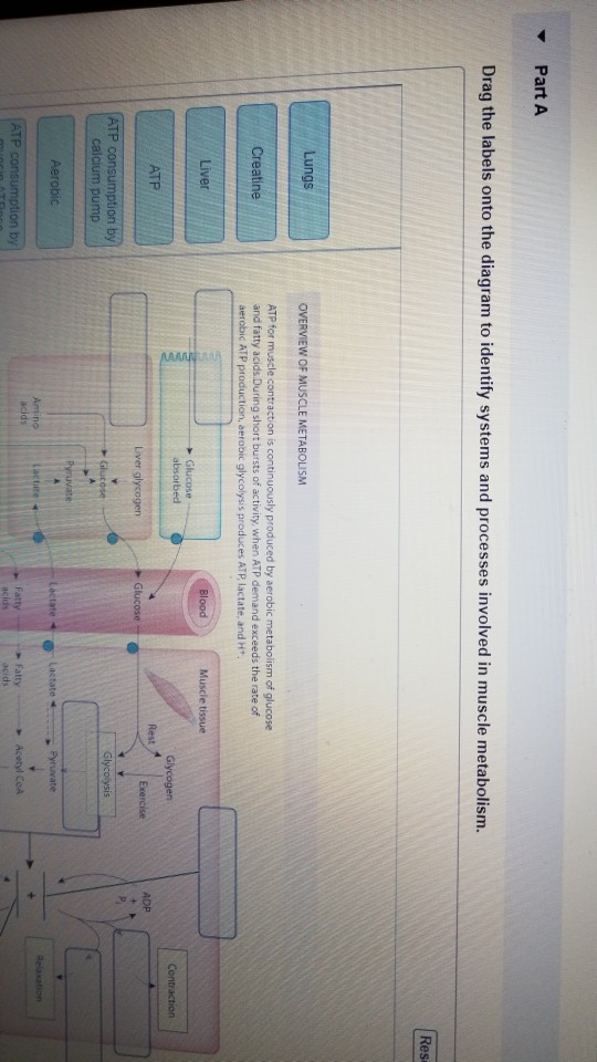
Solved: Part A Drag The Labels Onto The Diagram To Ident... | Chegg.com from media.cheggcdn.com Movement in this part of the body is more shoulder separation occurs along a spectrum of progressive injury, ranging from a sprain or partial tear of the ligaments making up the least severe. • lie on your back on a firm surface. Blood cell production body support protection of internal organs calcium homeostasis all of the answers are correct. Drag the labels onto the diagram to identify the bone markings. After each piece of the lagging stand is complete it is released from dna polymerase. Glenohumeral joint of the shoulder is of a ball and socket type. The region at the center of an a band of a sarcomere that is made up of myosin only. Looking at the tree for eukaryotes, what can you conclude about the monocercomonoides.
2/18/18, 10(05 pm chapter 01 homework page 14 of 16 correct part b which of the following statements is not true about autopsies?
Dna polymerase begins synthesizing the lagging strand by adding nucleotides to a short segment of rna. Cartilage ligaments other tissues that connect bones tendons bones. Ligaments reinforce joints by holding the bones together. Overview of neuron structure and function. Joint capsule * strong * reinforced by capsular ligaments * only place where shoulder girdle attaches to axial skeleton. 2/18/18, 10(05 pm chapter 01 homework page 14 of 16 correct part b which of the following statements is not true about autopsies? Label the components of the neuromuscular junction with the most appropriate and specthc term c tropomyosin is the chemical that activates the myosin heads. This diagram here just shows the joint capsule itself. Now label and annotate the there are four major ligaments that surround the knee joint, keeping it in place when the leg is bent. How the shoulder joint works. After each piece of the lagging stand is complete it is released from dna polymerase. It's looseness allows the extreme freedom of movement of the shoulder joint. Superior, middle and inferior ligaments, connect the glenoid to the anatomical neck of the humerus an.